- Simultaneously images cell protrusions and transport vesicles
- Up to three times faster than conventional methods
South Korean researchers have developed a microscopy technology capable of simultaneously observing the protrusions that living cancer cells extend to probe their surroundings and the tiny vesicles that ferry materials inside cells.
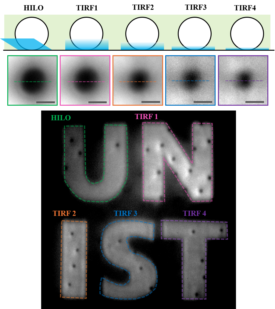
The Ulsan National Institute of Science and Technology (UNIST) announced Sunday that a research team led by Professor Park Jeong-hun of the Department of Biomedical Engineering had developed a "tunable total internal reflection fluorescence microscope" — or tunable TIRF — that adjusts the depth of illumination depending on the location within a cell.
Conventional fluorescence microscopes apply uniform lighting conditions across the entire field of view, making it difficult to image structures near the cell membrane and those at relatively greater heights with equal clarity at the same time. Observing cellular structures at different depths required repeated imaging sessions with adjusted lighting conditions each time.
To overcome these limitations, the research team added a digital micromirror device and a spatial filter to the microscope. The system works by adjusting the angles of millions of tiny mirrors to change the direction and depth at which light enters the sample.
The setup allows simultaneous observation of different depths — from shallow regions of 137 to 558 nanometers to the full height of a cell — within a single image.
The technology enables imaging up to twice as fast as conventional methods that alternate between two depths, and up to three times faster than those that capture three depths sequentially.
The research team used the microscope to observe human umbilical vein endothelial cells, capturing both cell membrane movement and the migration of internal vesicles in a single video.
In experiments on cervical cancer cells, the team successfully recorded the oscillation of cell protrusions and the movement of vesicles near the cell membrane simultaneously at 20 frames per second.
Image quality was also improved: by changing the illumination pattern of the micromirrors 1,440 times per second, the system reduced the streaks and blotches caused by light scattering.

Researchers can draw observation zones directly onto a cell image displayed on a tablet screen and specify the desired illumination depth for each zone; the microscope then automatically applies those conditions.
Another advantage is that the technology can be implemented by adding only a few components to an existing fluorescence microscope, eliminating the need to purchase expensive new equipment. Reducing the number of separate imaging passes also lowers the cells' cumulative exposure to light.
"We overcame the longstanding limitation of conventional microscopes — the requirement to apply a single lighting mode across the entire sample — through a new optical design," Park said. "The technology can be applied to studying a wide range of biological phenomena, including cell migration during cancer metastasis and wound healing, membrane transport, and bacterial infection, as well as to research on related treatments."
The findings were published in the Sept. 22 issue of the international journal Laser & Photonics Reviews.
nbgkoo@heraldcorp.com