Study tracking 226 patients shows stable foot function after free perforator flap reconstruction
Professor Lee Kyung-tae vows to refine surgical techniques with patient quality of life in mind
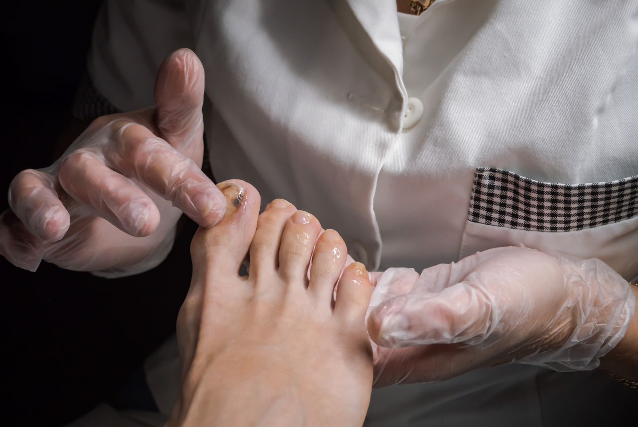
Patients who undergo precise microsurgical reconstruction tailored to the condition of their foot after melanoma removal can maintain good daily function with minimal pain and reduced loss of mobility, according to new research.
Foot melanoma is a cancer arising from pigment-producing cells anywhere on the foot — including the toes, the top of the foot and the sole. Treatment requires wide excision of the tumor followed by reconstruction of the resulting defect. The challenge is that the foot has limited skin available to close a wound, and its role in bearing the body's full weight makes reconstruction especially demanding. The sole and heel must withstand constant daily pressure and friction, while the toes need to be reconstructed thinly and naturally to allow comfortable shoe wear and walking.
To address these challenges, a research team from Samsung Medical Center — comprising plastic surgeons Lee Kyung-tae, Moon Gu-hyeon and Kim Jin-a, along with dermatologist Lee Dong-yun — investigated the potential of free perforator flap surgery and published their findings in a recent issue of the international journal Plastic and Reconstructive Surgery.
The team analyzed outcomes for 226 patients who underwent immediate free perforator flap reconstruction following foot melanoma removal between 2005 and 2023. The procedure involves harvesting skin tissue containing blood vessels from the thigh, back or groin and connecting it to blood vessels in the foot under a microscope.
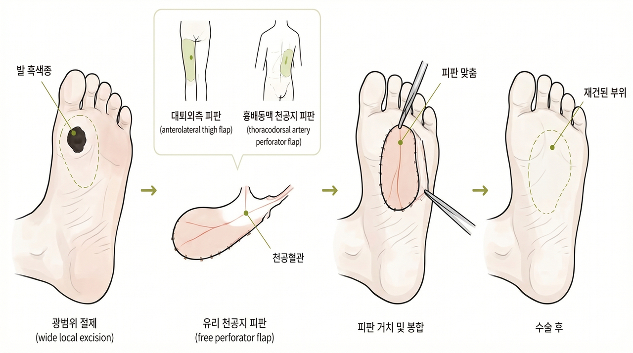
Because donor tissue from other parts of the body tends to be thicker than the foot's own skin, there is a risk of protrusion or displacement at the surgical site after the procedure. The research team overcame this by thinning the transplanted tissue to match the depth of each defect and by selecting different donor sites and blood vessels depending on the location — whether the toe, sole or heel.
According to the research, graft failure occurred in only five of the 226 patients, a rate of 2.2 percent, and most complications were minor, amounting to delayed wound healing. Among the 70 patients for whom long-term follow-up was possible, foot function was assessed using a scoring index that measures pain, difficulty using the foot, and limitations in daily activities and movement. At a median follow-up of 17 months after surgery, patients scored an average of 7.9 out of 100 — a low score indicating minimal pain and functional limitation.
Patients who required complex microsurgery because of large defects showed no significant difference in postoperative foot function scores compared with those who had smaller defects repaired through skin grafting or local flap procedures. The research team said this demonstrated that even extensive defects can be reconstructed with precision to minimize pain and disruption to daily life.

"For patients with foot melanoma, pain after surgery and the ability to carry on with daily life are just as important as removing the tumor itself," Lee said. "This study confirms that tailored reconstruction — one that accounts for the location of the defect and the patient's future walking function — can deliver treatment that also addresses quality of life. We will continue to push forward in refining our surgical techniques."
kty@heraldcorp.com