- DGIST and POSTECH create 3D muscle tissue from real human cells
- Aging model shows thinner fibers, weaker contractions and higher inflammation, offering an alternative to animal testing
South Korean researchers have developed a three-dimensional artificial muscle that replicates the aging process of human muscle tissue, using cells donated by a 17-year-old and a 68-year-old. The advance is expected to open the door to analyzing age-related muscle decline and evaluating treatments for sarcopenia without relying on animal experiments.
The Daegu Gyeongbuk Institute of Science and Technology (DGIST) said Thursday that a research team led by Professor Kim Min-seok of its Department of New Biology, working jointly with a team led by Professor Cho Dong-woo of POSTECH's Department of Mechanical Engineering, had developed a "biomimetic 3D skeletal muscle micro-tissue model" that simultaneously reproduces human muscle aging and the accompanying loss of contractile force.
Skeletal muscle is a core tissue that attaches to bone, drives physical movement and regulates energy metabolism. Sarcopenia — the progressive loss of muscle mass and strength with age — is widely regarded as a leading cause of reduced physical activity and diminished quality of life in older adults.
Existing research approaches, however, have faced significant limitations. Animal models differ substantially from humans in biological terms, while conventional two-dimensional cell cultures grown in flat dishes struggle to replicate the three-dimensional structure and contractile function of real muscle tissue.
To overcome these constraints, the research team used muscle cells obtained from male donors aged 17 and 68.
The cells were mixed with a biomaterial called decellularized extracellular matrix, which serves as a structural scaffold mimicking real tissue, and then cultured inside a microscale mold fabricated by 3D printing. The process produced a three-dimensional micro-tissue in which the muscle cells aligned in a uniform direction, closely resembling the architecture of actual human muscle.
The artificial muscle built from the 68-year-old donor's cells showed thinner muscle fibers and a lower degree of cell fusion than the tissue derived from the 17-year-old donor. Inflammation-related markers were also elevated.
Mitochondrial function, which is responsible for cellular energy production, was impaired. Calcium responses that trigger muscle contraction were slower, and the actual contractile force generated under electrical stimulation was markedly reduced.
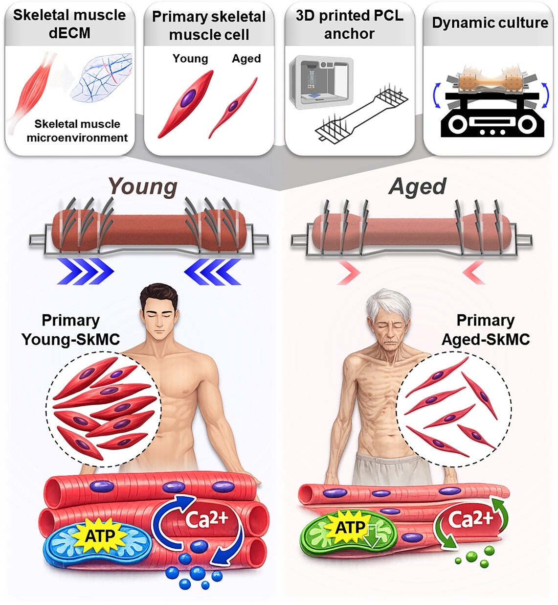
Through these findings, the research team confirmed that both the structural changes and the physiological decline characteristic of human muscle aging could be observed simultaneously within a single artificial tissue construct.
The technology is particularly significant because it goes beyond simply observing changes in cell shape or gene expression — it allows researchers to directly measure how forcefully the muscle actually contracts.
The team expects the model to serve as a comprehensive evaluation tool: candidate treatments for sarcopenia can be applied to the artificial muscle, and researchers can then assess changes in muscle fiber morphology, inflammatory responses, mitochondrial function and improvements in contractile force all at once.
The platform's potential as a next-generation drug development tool that could replace animal testing has also drawn attention.
The US Food and Drug Administration (FDA) has recently been updating its regulatory framework to allow non-clinical test data obtained through methods other than animal experiments to be used in drug development and approval processes. Against that backdrop, the value of three-dimensional artificial tissues that reflect real human physiological characteristics is growing.
"This research is highly significant in that it vividly reproduces aging-related changes using human-derived cells and lays the groundwork for evaluating both cellular-level changes and actual contractile function together," Kim said. Kim added that the team would develop the model into a useful research tool for identifying the causes of muscle aging and accurately verifying the efficacy of treatment candidates.
The findings were selected as a cover paper in the international journal Small Science.
nbgkoo@heraldcorp.com