According to the latest statistics from the National Cancer Information Center, malignant melanoma accounts for about 10 percent of all skin cancers in South Korea, with a five-year survival rate exceeding 90 percent. While that figure is high compared with other types of skin cancer, it underscores the importance of recognizing symptoms early and responding quickly. Particularly close observation is needed when the condition develops in areas that are easy to overlook, such as the skin, toenails, fingernails or the soles of the feet.
The American Cancer Society offers the ABCDE framework for identifying malignant melanoma. A stands for asymmetry: normal moles and other benign lesions tend to be symmetrical. Malignant melanoma, however, is irregular in shape and asymmetrical in more than 80 percent of cases. An uneven, map-like outline is a warning sign.
B stands for border irregularity: the edges are often uneven, jagged and poorly defined. While the borders of a normal mole are relatively clear and rounded, malignant melanoma has blurred or serrated edges in as many as 75 percent of cases.
C stands for color variation: rather than a single uniform color, the lesion often displays a mix of shades. In addition to brown and black, red, blue and white may appear together, and more than 60 percent of malignant melanomas show this multicolored characteristic.
D stands for diameter: most malignant melanomas are 6 millimeters or more in diameter. That is roughly the size of a pencil eraser, and any mole larger than that which continues to grow or change shape should be evaluated by a specialist.
E stands for evolution: a mole that changes in size, shape, color or elevation over time is more likely to be malignant melanoma. Cases in which a lesion changes by more than 20 percent in a year, or grows by more than 2 millimeters in a year, warrant careful monitoring.
Malignant melanoma arising on toenails, fingernails or the soles of the feet can present differently from ordinary moles and requires extra vigilance. A key symptom is the appearance of a vertical black or brown streak beneath a fingernail or toenail. If the streak is 3 millimeters or wider, has uneven coloring, or gradually thickens and spreads over time, malignant melanoma should be suspected. Black pigment may also spread to the skin surrounding the nail — a phenomenon known as Hutchinson's sign — which raises the probability of malignant melanoma to more than 70 percent. Bleeding or ulceration beneath a fingernail or toenail also warrants attention.
A diagnosis of melanoma in the fingernails or toenails can in the worst cases require partial amputation of the hand or foot. A South Korean research team has now published findings showing that amputation is not always necessary for nail melanoma patients. A team led by Lee Kyung-tae, a plastic surgery professor at Samsung Medical Center, and resident Lee Eun-song, working with the dermatology department, announced results demonstrating that function-preserving surgery without amputation is possible even in patients with deeply invasive subungual melanoma, provided the bone is not involved.
Melanoma is a skin cancer originating in melanocytes. In its early stages it can resemble an ordinary mole or bruise, making it easy to overlook. Acral melanoma — the form that develops at the body's extremities, including the fingers and toes — is rare but known to progress rapidly and carry a poor prognosis.
Nail melanoma has long been treated by amputating the entire affected joint, given the high likelihood that cancer cells would invade the bone, and amputation has been considered the standard of care. The problem is the significant functional and psychological burden amputation places on patients. Function-preserving surgery to save the digit was attempted as an alternative, but wide variation in outcomes — driven by individual surgeons' subjective judgment — kept it from being considered a first-line option.
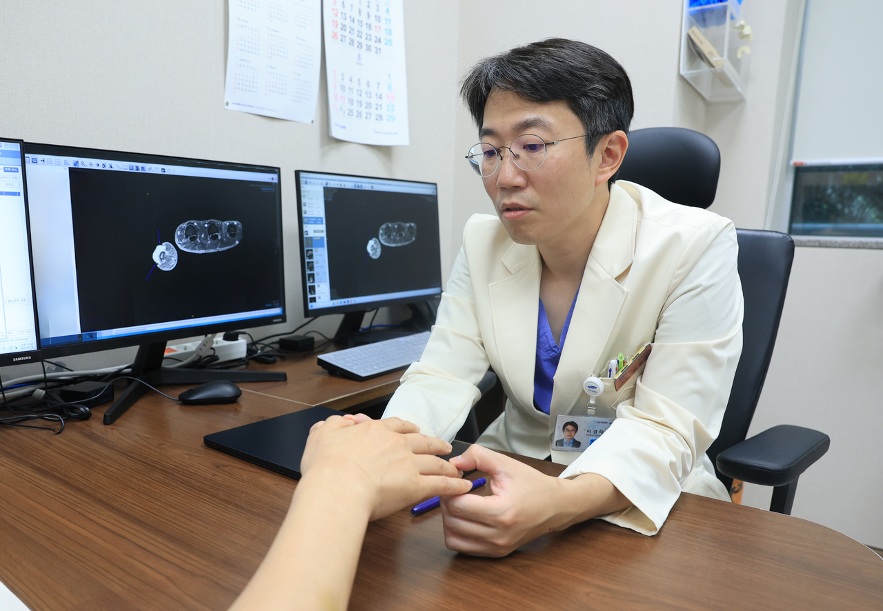
To address that limitation, the research team added an objective protocol: pre-operative MRI to confirm whether bone invasion was present before surgery. Applying this MRI-based function-preserving surgical protocol to 27 patients with invasive nail melanoma between 2021 and 2025, the team demonstrated that complete tumor removal is achievable without amputation.
The key is using precision MRI before surgery to objectively confirm whether bone invasion has actually occurred. When MRI showed no bone involvement, the team proceeded — regardless of tumor thickness — by precisely excising only the cancerous tissue and then transplanting a thin skin-and-microvascular flap harvested from the groin to reconstruct the digit, preserving both its shape and function. The technique is known as the superthin SCIP flap.
The team said every patient whose MRI was read as showing no bone invasion was confirmed by biopsy to have no bone involvement, establishing the high clinical reliability of MRI in this setting.
Treatment outcomes were also strong, the team said. Analysis of the follow-up period showed a local recurrence rate of zero — not a single case — while the two-year disease-free survival rate was 81.6 percent and the two-year locoregional recurrence-free survival rate was 94.4 percent. The team said these results demonstrate oncological safety comparable to conventional amputation.
"Avoiding unnecessary amputation and improving patients' quality of life is a critical responsibility for clinicians," said Lee Kyung-tae, the plastic surgery professor at Samsung Medical Center who led the research. "This study will serve as a milestone showing that, when backed by pre-operative imaging and advanced reconstructive techniques, patients' quality of life can be dramatically preserved without amputation."

Meanwhile, the research team noted that the study was conducted at a single institution and involved only Asian patients, and said it plans to carry out larger, multi-center studies including more diverse ethnic groups to confirm the broader applicability of the protocol. The findings were published in the latest issue of the European Journal of Surgical Oncology (impact factor: 2.9).
kty@heraldcorp.com